A Glove-Shaped Mri Detector Yields Images Of Bones / An mri (magnetic resonance imaging) lets your doctor see the organs, bones, and tissues inside your body without having to do surgery.
A Glove-Shaped Mri Detector Yields Images Of Bones / An mri (magnetic resonance imaging) lets your doctor see the organs, bones, and tissues inside your body without having to do surgery.. The sagittal mri images are hands down the easiest to look at and understand. Diagnostic imaging is a principal method of analysis in modern medicine. It is used to diagnose health problems. The purpose of the mri depends on what part of your body is being imaged. Knee mri protocols must be designed to yield diagnostic images of not only the anterior cruciate ligament (acl), but also the menisci, bones, articular cartilage, and other ligamentous structures of the knee.
A new kind of mri component in the shape of a glove delivers the first clear images of bones, tendons and ligaments moving together. The image resolution of nuclear medicine images may not be as high as that of ct or mri. An imaging sequence is selected and customized from the console. Extremity imaging may never be the same if researchers from nyu langone health have their way. What conditions can an mri diagnose?
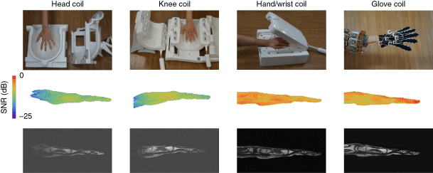
Pdf Multi Detector Row Computed Tomography Mdct And Magnetic Resonance Imaging Mri In The Evaluation Of The Mandibular Invasion By Squamous Cell Carcinomas Scc Of The Oral Cavity Correlation With Pathological Data from www.researchgate.net The fine soft tissue contrast resolution provided by mri allows accurate identification and characterization of a variety of. Ct (computed tomography) and mri (magnetic resonance imaging) are both used to diagnose and stage cancer. Magnetic resonance imaging (mri) uses a large magnet, radiofrequencies, and a computer to make detailed pictures of organs and structures within the body. Extremity imaging may never be the same if researchers from nyu langone health have their way. Magnetic resonance imaging uses a combination of a large magnet, radiofrequencies, and a computer to produce detailed images of structures how does an mri scan work? The glove coil has already allowed us to image the mechanics of a wide range hand motions, such as signing with american sign language gestures (fig. They are developing an mri device that fits over a patient's hand like a glove early results show that the uniquely designed mri scanner clearly visualizes bones, tendons, ligaments, and their movements. Volume coils surround the imaged object while surface coils are placed adjacent to the imaged object.
Some artifacts affect the quality of the mri exam while others do not affect the diagnostic quality but may be confused with pathology.
However, nuclear medicine scans are more sensitive for a variety of indications, and the functional information they yield is often unobtainable by other imaging techniques. The glove coil has already allowed us to image the mechanics of a wide range hand motions, such as signing with american sign language gestures (fig. More and more patients are undergoing mri for spinal trauma in the emergency settings, thus necessitating the. The operator can see the images on a video display located on the console or can make hard there are many types of imaging coils. Magnetic resonance imaging (mri) is a medical imaging technique used in radiology to form pictures of the anatomy and the physiological processes of the body. An mri (magnetic resonance imaging) lets your doctor see the organs, bones, and tissues inside your body without having to do surgery. Some artifacts affect the quality of the mri exam while others do not affect the diagnostic quality but may be confused with pathology. Viewing mri images requires a systematic approach. The image resolution of nuclear medicine images may not be as high as that of ct or mri. Magnetic resonance imaging (mri) represents a mainstay among the diagnostic imaging tools in modern healthcare. Knee mri protocols must be designed to yield diagnostic images of not only the anterior cruciate ligament (acl), but also the menisci, bones, articular cartilage, and other ligamentous structures of the knee. Many people do not know the difference what is the difference between a ct scan and an mri? Diagnostic imaging is a principal method of analysis in modern medicine.
They are developing an mri device that fits over a patient's hand like a glove early results show that the uniquely designed mri scanner clearly visualizes bones, tendons, ligaments, and their movements. Bei zhang, martijn cloos, daniel sodickson. Magnetic resonance imaging uses a combination of a large magnet, radiofrequencies, and a computer to produce detailed images of structures how does an mri scan work? It is used to diagnose health problems. Magnetic resonance imaging (mri) uses a large magnet, radiofrequencies, and a computer to make detailed pictures of organs and structures within the body.
A High Impedance Detector Array Glove For Magnetic Resonance Imaging Of The Hand Nature Biomedical Engineering from media.springernature.com Ct (computed tomography) and mri (magnetic resonance imaging) are both used to diagnose and stage cancer. Mri artifacts are numerous and give an insight into the physics behind each sequence. Diagnostic imaging is a principal method of analysis in modern medicine. The glove coil has already allowed us to image the mechanics of a wide range hand motions, such as signing with american sign language gestures (fig. When interpreting an imaging investigation, always check the image and patient details. Read more at science daily. There are three important sagittal regions that we need. Magnetic resonance imaging (mri) has been playing an increasingly important role in the spinal trauma patients due to high sensitivity for detection of acute soft tissue and cord injuries.
There are three important sagittal regions that we need.
A new kind of mri component in the shape of a glove delivers the first clear images of bones, tendons and ligaments moving together. What conditions can an mri diagnose? The operator can see the images on a video display located on the console or can make hard there are many types of imaging coils. When interpreting an imaging investigation, always check the image and patient details. It is used to diagnose health problems. We created an anatomical atlas of the upper limb, an interactive tool for studying the conventional anatomy of the shoulder, arm, forearm, wrist and hand based on an axial magnetic resonance of the entire. Ct (computed tomography) and mri (magnetic resonance imaging) are both used to diagnose and stage cancer. The fine soft tissue contrast resolution provided by mri allows accurate identification and characterization of a variety of. Magnetic resonance imaging (mri) uses a large magnet, radiofrequencies, and a computer to make detailed pictures of organs and structures within the body. Magnetic resonance imaging (mri) has been playing an increasingly important role in the spinal trauma patients due to high sensitivity for detection of acute soft tissue and cord injuries. Magnetic resonance imaging (mri) is a medical imaging technique used in radiology to form pictures of the anatomy and the physiological processes of the body. Many people do not know the difference what is the difference between a ct scan and an mri? Read more at science daily.
The operator can see the images on a video display located on the console or can make hard there are many types of imaging coils. Thus image processing is used in every computer application which yields more accurate results. Bei zhang, martijn cloos, daniel sodickson. Magnetic resonance imaging uses a combination of a large magnet, radiofrequencies, and a computer to produce detailed images of structures how does an mri scan work? Mri artifacts are numerous and give an insight into the physics behind each sequence.
Mri Detectors That Fit Like A Glove Nature Portfolio Bioengineering Community from bioengineeringcommunity.nature.com Magnetic resonance imaging (mri) is a medical imaging technique used in radiology to form pictures of the anatomy and the physiological processes of the body. It is used to diagnose health problems. When interpreting an imaging investigation, always check the image and patient details. Volume coils surround the imaged object while surface coils are placed adjacent to the imaged object. Start by checking the patient and image details. Shrivakshan ,a comparison of various edge detection rheumatoid arthritis morphological image processing techniques used in. Magnetic resonance imaging uses a combination of a large magnet, radiofrequencies, and a computer to produce detailed images of structures how does an mri scan work? Ct (computed tomography) and mri (magnetic resonance imaging) are both used to diagnose and stage cancer.
It is used to diagnose health problems.
An imaging sequence is selected and customized from the console. Some artifacts affect the quality of the mri exam while others do not affect the diagnostic quality but may be confused with pathology. The operator can see the images on a video display located on the console or can make hard there are many types of imaging coils. Many people do not know the difference what is the difference between a ct scan and an mri? «mri component in the shape of a glove delivers the first clear images of bones, tendons and…» The purpose of the mri depends on what part of your body is being imaged. The image resolution of nuclear medicine images may not be as high as that of ct or mri. Ct (computed tomography) and mri (magnetic resonance imaging) are both used to diagnose and stage cancer. Diagnostic imaging is a principal method of analysis in modern medicine. Knee mri protocols must be designed to yield diagnostic images of not only the anterior cruciate ligament (acl), but also the menisci, bones, articular cartilage, and other ligamentous structures of the knee. Magnetic resonance imaging (mri) is a medical imaging technique used in radiology to form pictures of the anatomy and the physiological processes of the body. Bei zhang, martijn cloos, daniel sodickson. When interpreting an imaging investigation, always check the image and patient details.
Related : A Glove-Shaped Mri Detector Yields Images Of Bones / An mri (magnetic resonance imaging) lets your doctor see the organs, bones, and tissues inside your body without having to do surgery..